Copyright
©The Author(s) 2015.
World J Gastroenterol. Feb 21, 2015; 21(7): 2229-2235
Published online Feb 21, 2015. doi: 10.3748/wjg.v21.i7.2229
Published online Feb 21, 2015. doi: 10.3748/wjg.v21.i7.2229
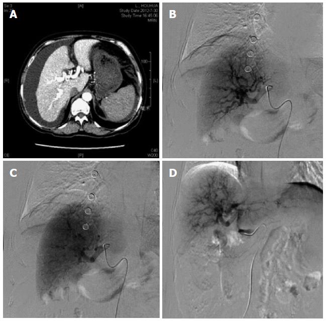
Figure 1 Contrast-enhanced computed tomography revealed an earlier enhancement of the portal vein compared with the superior mesenteric vein during the arterial phase.
Digital subtraction angiography indicated that there was rapid filling through the fistula into the portal vein. A: Early enhancement of the portal branches in hepatic arterial-phase; B, C: The hepatic argiography; D: The fistula reduction after embolization.
- Citation: Zhang DY, Weng SQ, Dong L, Shen XZ, Qu XD. Portal hypertension induced by congenital hepatic arterioportal fistula: Report of four clinical cases and review of the literature. World J Gastroenterol 2015; 21(7): 2229-2235
- URL: https://www.wjgnet.com/1007-9327/full/v21/i7/2229.htm
- DOI: https://dx.doi.org/10.3748/wjg.v21.i7.2229


