Copyright
©The Author(s) 2015.
World J Gastroenterol. Feb 21, 2015; 21(7): 2220-2224
Published online Feb 21, 2015. doi: 10.3748/wjg.v21.i7.2220
Published online Feb 21, 2015. doi: 10.3748/wjg.v21.i7.2220
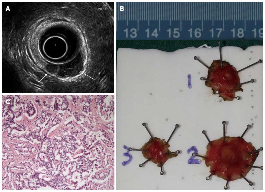
Figure 1 Ultrasound image, surgical specimens and pathological image of the carcinoids.
A: Transrectal ultrasound image showing two hypoechoic nodules (arrow), 0.5 cm and 0.72 cm in maximum diameter (lesions 1 and 2, respectively), confined to the submucosal layer of the rectal wall; B: Surgical specimens of the three lesions (two carcinoids, specimen 1 and 2; and one scar site with residual tumor, specimen 3); C: The pathological image revealing neuroendocrine tumor cells within the submucosal layer (hematoxylin and eosin, × 100).
- Citation: Zhou JL, Lin GL, Zhao DC, Zhong GX, Qiu HZ. Resection of multiple rectal carcinoids with transanal endoscopic microsurgery: Case report. World J Gastroenterol 2015; 21(7): 2220-2224
- URL: https://www.wjgnet.com/1007-9327/full/v21/i7/2220.htm
- DOI: https://dx.doi.org/10.3748/wjg.v21.i7.2220


