Copyright
©The Author(s) 2015.
World J Gastroenterol. Feb 21, 2015; 21(7): 2159-2168
Published online Feb 21, 2015. doi: 10.3748/wjg.v21.i7.2159
Published online Feb 21, 2015. doi: 10.3748/wjg.v21.i7.2159
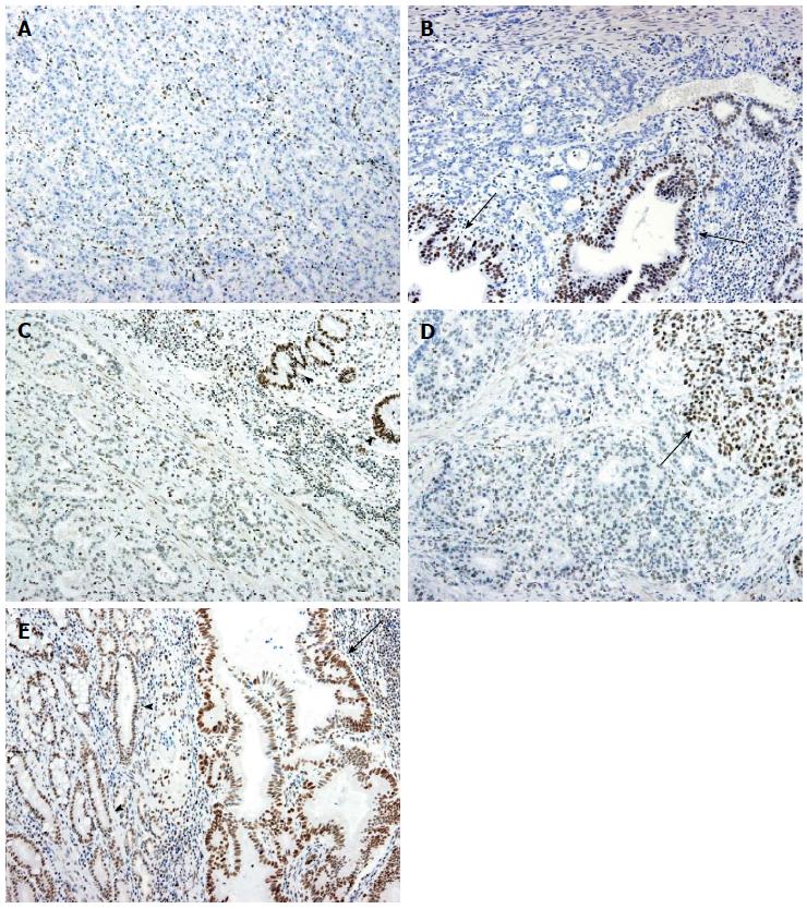
Figure 2 Immunohistochemistry for ARID1A.
A: Homogeneous loss of expression. All the tumor cells show no expression, whereas the stromal cells retain the nuclear expression of ARID1A; B: Heterogeneous loss of expression. Most of the tumor cells show no expression, whereas some of the gland-forming tumor cells retain nuclear expression (arrows); C: Homogeneously weak expression. The tumor cells show the reduced expression of ARID1A. Non-neoplastic gastric glandular cells retain the expression (arrowheads); D: Heterogeneously weak expression. Most of the tumor cells exhibit reduced expression, but a subset of tumor cells retain nuclear expression (arrow); E: Retained expression: Tumor cells (arrow) show strong nuclear ARID1A expression, similar to non-neoplastic glandular cells (arrowheads).
- Citation: Inada R, Sekine S, Taniguchi H, Tsuda H, Katai H, Fujiwara T, Kushima R. ARID1A expression in gastric adenocarcinoma: Clinicopathological significance and correlation with DNA mismatch repair status. World J Gastroenterol 2015; 21(7): 2159-2168
- URL: https://www.wjgnet.com/1007-9327/full/v21/i7/2159.htm
- DOI: https://dx.doi.org/10.3748/wjg.v21.i7.2159


