Copyright
©The Author(s) 2015.
World J Gastroenterol. Feb 14, 2015; 21(6): 1915-1926
Published online Feb 14, 2015. doi: 10.3748/wjg.v21.i6.1915
Published online Feb 14, 2015. doi: 10.3748/wjg.v21.i6.1915
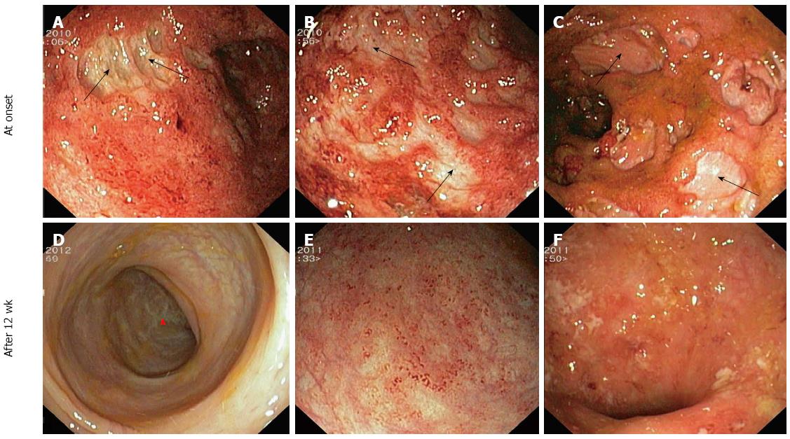
Figure 5 Presence of holes in the mucosa with exposure of the underlying muscular layer (black arrows), surrounded by granular and spontaneously bleeding zones, is clearly evident (panels A to C).
After a three-month washout period from any therapy for the primary disease, the healing process with white scars (red arrow) was observed in the only patient who underwent a cycle with rituximab (panel D), whilst a slight or no improvement was observed in the other two representative cases.
- Citation: Ciccocioppo R, Racca F, Paolucci S, Campanini G, Pozzi L, Betti E, Riboni R, Vanoli A, Baldanti F, Corazza GR. Human cytomegalovirus and Epstein-Barr virus infection in inflammatory bowel disease: Need for mucosal viral load measurement. World J Gastroenterol 2015; 21(6): 1915-1926
- URL: https://www.wjgnet.com/1007-9327/full/v21/i6/1915.htm
- DOI: https://dx.doi.org/10.3748/wjg.v21.i6.1915


