Copyright
©The Author(s) 2015.
World J Gastroenterol. Feb 7, 2015; 21(5): 1580-1587
Published online Feb 7, 2015. doi: 10.3748/wjg.v21.i5.1580
Published online Feb 7, 2015. doi: 10.3748/wjg.v21.i5.1580
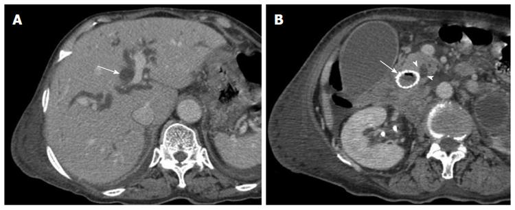
Figure 2 Images from an 81-year-old man with bladder cancer with multiple metastases.
A: Computed tomographic (CT) scan obtained 18 d after stent placement showing significant dilatation of the intrahepatic bile duct (long arrow); B: CT scan obtained 18 d after stent placement at the level of the ampulla of Vater (AOV) showing significant dilatation of the common bile duct due to the compression of the inserted stent (long arrow) to the AOV (arrowheads).
- Citation: Kim JW, Jeong JB, Lee KL, Kim BG, Ahn DW, Lee JK, Kim SH. Comparison between uncovered and covered self-expandable metal stent placement in malignant duodenal obstruction. World J Gastroenterol 2015; 21(5): 1580-1587
- URL: https://www.wjgnet.com/1007-9327/full/v21/i5/1580.htm
- DOI: https://dx.doi.org/10.3748/wjg.v21.i5.1580


