Copyright
©The Author(s) 2015.
World J Gastroenterol. Feb 7, 2015; 21(5): 1554-1566
Published online Feb 7, 2015. doi: 10.3748/wjg.v21.i5.1554
Published online Feb 7, 2015. doi: 10.3748/wjg.v21.i5.1554
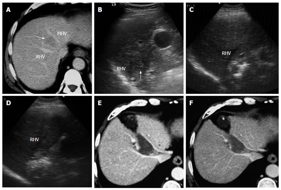
Figure 5 A 54-year-old man with hepatocellular carcinoma.
A: Contrast-enhanced transverse computed tomography (CT) image shows a 2.6-cm tumor (arrow) in segment VIII between the middle hepatic vein (MHV) and the right hepatic vein (RHV); B: Intercostal ultrasound shows that the tumor (arrow) is located next to RHV; C: Intercostal ultrasound shows that the tumor is treated by ultrasound-guided RF ablation. Two bipolar electrodes are inserted parallel to the RHV; D: Intercostal ultrasound immediately after RF ablation shows the hepatic veins remain normal; E: Contrast-enhanced transverse CT image obtained 1 mo later shows that a coagulation area is surrounded by hepatic veins with no enhancement. No injury to the large vessels occurred in this patient; F: Contrast-enhanced transverse CT image obtained 5 mo later shows a coagulation area without viability.
- Citation: Yang W, Yan K, Wu GX, Wu W, Fu Y, Lee JC, Zhang ZY, Wang S, Chen MH. Radiofrequency ablation of hepatocellular carcinoma in difficult locations: Strategies and long-term outcomes. World J Gastroenterol 2015; 21(5): 1554-1566
- URL: https://www.wjgnet.com/1007-9327/full/v21/i5/1554.htm
- DOI: https://dx.doi.org/10.3748/wjg.v21.i5.1554


