Copyright
©The Author(s) 2015.
World J Gastroenterol. Feb 7, 2015; 21(5): 1554-1566
Published online Feb 7, 2015. doi: 10.3748/wjg.v21.i5.1554
Published online Feb 7, 2015. doi: 10.3748/wjg.v21.i5.1554
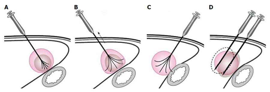
Figure 1 Diagram for ablation of a tumor near the bowel.
A: The multiple-tined electrode was inserted into the tumor perpendicular to the bowel wall, and the needle was expanded to 3 cm; B: When expanding the radiofrequency ablation (RFA) prongs from 3 cm to 4-5 cm to ablate a tumor area near the bowel, the RFA electrode was retracted slightly to draw the tumor away from the bowel. While fixing the electrode shaft in this position, the movable hub was pushed to expand the prongs; C: If the RFA electrode was inserted into the tumor parallel to the bowel wall, there is a higher risk for bowel injury when deploying the electrode tines; D: With a mono-polar electrode, the puncture direction was parallel to the wall of the adjacent bowel.
- Citation: Yang W, Yan K, Wu GX, Wu W, Fu Y, Lee JC, Zhang ZY, Wang S, Chen MH. Radiofrequency ablation of hepatocellular carcinoma in difficult locations: Strategies and long-term outcomes. World J Gastroenterol 2015; 21(5): 1554-1566
- URL: https://www.wjgnet.com/1007-9327/full/v21/i5/1554.htm
- DOI: https://dx.doi.org/10.3748/wjg.v21.i5.1554


