Copyright
©The Author(s) 2015.
World J Gastroenterol. Dec 28, 2015; 21(48): 13542-13547
Published online Dec 28, 2015. doi: 10.3748/wjg.v21.i48.13542
Published online Dec 28, 2015. doi: 10.3748/wjg.v21.i48.13542
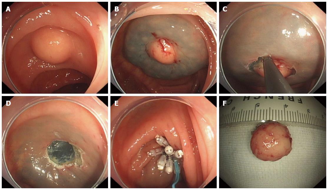
Figure 2 Endoscopic submucosal dissection procedure.
A: A 1.0 cm × 1.2 cm hemispherical submucosal lesion which had a hard texture, fixed, smooth surface, was observed in the ileocecal junction; B: Saline solution with a small amount of epinephrine and indigo carmine was injected beneath the lesion, and resulted in good elevation; C: Complete dissection of the lesion after circumferential incision; D: The lesion was completely removed without any bleeding or perforation; E: Purse-string suture with metallic clips and endoloop; F: Dissected specimen was sent for pathologic examination.
- Citation: Take I, Shi Q, Qi ZP, Cai SL, Yao LQ, Zhou PH, Zhong YS. Endoscopic resection of colorectal granular cell tumors. World J Gastroenterol 2015; 21(48): 13542-13547
- URL: https://www.wjgnet.com/1007-9327/full/v21/i48/13542.htm
- DOI: https://dx.doi.org/10.3748/wjg.v21.i48.13542


