Copyright
©The Author(s) 2015.
World J Gastroenterol. Dec 28, 2015; 21(48): 13542-13547
Published online Dec 28, 2015. doi: 10.3748/wjg.v21.i48.13542
Published online Dec 28, 2015. doi: 10.3748/wjg.v21.i48.13542
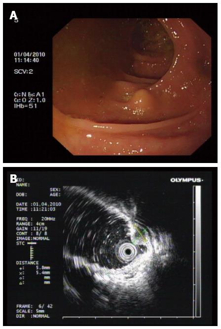
Figure 1 Preoperative examination.
A: A 0.8 cm submucosal lesion was observed 60 cm from the anal verge (ascending colon). The mass was sessile, the mucosal surface was smooth, with non-necrotic erosion, and was non-circumferential. B: Ultrasonographic examination revealed a 0.5 cm hypoechoic submucosal lesion without muscularis propria involvement.
- Citation: Take I, Shi Q, Qi ZP, Cai SL, Yao LQ, Zhou PH, Zhong YS. Endoscopic resection of colorectal granular cell tumors. World J Gastroenterol 2015; 21(48): 13542-13547
- URL: https://www.wjgnet.com/1007-9327/full/v21/i48/13542.htm
- DOI: https://dx.doi.org/10.3748/wjg.v21.i48.13542


