Copyright
©The Author(s) 2015.
World J Gastroenterol. Dec 28, 2015; 21(48): 13418-13431
Published online Dec 28, 2015. doi: 10.3748/wjg.v21.i48.13418
Published online Dec 28, 2015. doi: 10.3748/wjg.v21.i48.13418
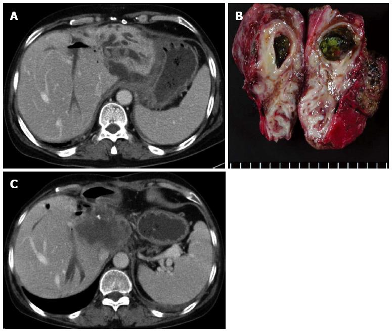
Figure 4 Forty-six years old, female was admitted for cholangitis.
A: Abdominal CT demonstrated that multiple calcified stones are present in the lateral segment of the left lobe; B: Liver, left lobectomy was performed, and there was no cancerous lesion; C: Abdominal computed tomography taken 14 mo after resection demonstrated development of cholangiocarcinoma in the caudate lobe of the liver.
- Citation: Kim HJ, Kim JS, Joo MK, Lee BJ, Kim JH, Yeon JE, Park JJ, Byun KS, Bak YT. Hepatolithiasis and intrahepatic cholangiocarcinoma: A review. World J Gastroenterol 2015; 21(48): 13418-13431
- URL: https://www.wjgnet.com/1007-9327/full/v21/i48/13418.htm
- DOI: https://dx.doi.org/10.3748/wjg.v21.i48.13418


