Copyright
©The Author(s) 2015.
World J Gastroenterol. Dec 14, 2015; 21(46): 13140-13151
Published online Dec 14, 2015. doi: 10.3748/wjg.v21.i46.13140
Published online Dec 14, 2015. doi: 10.3748/wjg.v21.i46.13140
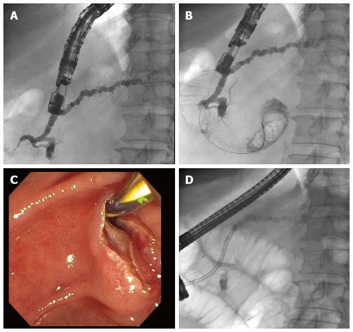
Figure 1 Patient with dilated pancreatic duct, recurrent pancreatitis, and pancreas divisum in whom endoscopic retrograde cholangiopancreatography has failed.
A: After endoscopic ultrasonography-guided puncture and administration of contrast medium, imaging of the pancreatic duct under fluoroscopic control shows there is almost no outflow of contrast medium out of the minor papilla; B: The guide wire is advanced into the pancreatic duct and through and out of the minor papilla; C: After changing to a duodenoscope, the guide wire was identified and an extending papillotomy was performed using a conventional technique; D: An 11.5-Fr. prosthesis is placed into the minor papilla, with subsequent adequate outflow of contrast medium.
- Citation: Will U, Reichel A, Fueldner F, Meyer F. Endoscopic ultrasonography-guided drainage for patients with symptomatic obstruction and enlargement of the pancreatic duct. World J Gastroenterol 2015; 21(46): 13140-13151
- URL: https://www.wjgnet.com/1007-9327/full/v21/i46/13140.htm
- DOI: https://dx.doi.org/10.3748/wjg.v21.i46.13140


