Copyright
©The Author(s) 2015.
World J Gastroenterol. Dec 14, 2015; 21(46): 13020-13029
Published online Dec 14, 2015. doi: 10.3748/wjg.v21.i46.13020
Published online Dec 14, 2015. doi: 10.3748/wjg.v21.i46.13020
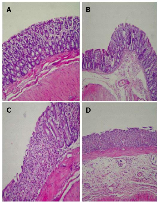
Figure 3 Histologic images of rat colon.
A: Normal control; B: Kefir-control, mild acute inflammation in Lamina propria; C: Colitis, extensive ulceration of the surface epithelium and crypt loss; D: Kefir-colitis, ulceration of the epithelial surface and crypt loss, mild inflammation, and moderate submucosal edema (HE, magnification × 100).
- Citation: Senol A, Isler M, Sutcu R, Akin M, Cakir E, Ceyhan BM, Kockar MC. Kefir treatment ameliorates dextran sulfate sodium-induced colitis in rats. World J Gastroenterol 2015; 21(46): 13020-13029
- URL: https://www.wjgnet.com/1007-9327/full/v21/i46/13020.htm
- DOI: https://dx.doi.org/10.3748/wjg.v21.i46.13020


