Copyright
©The Author(s) 2015.
World J Gastroenterol. Dec 7, 2015; 21(45): 12778-12786
Published online Dec 7, 2015. doi: 10.3748/wjg.v21.i45.12778
Published online Dec 7, 2015. doi: 10.3748/wjg.v21.i45.12778
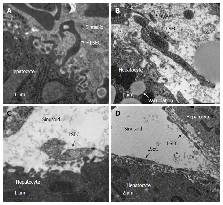
Figure 5 Transmission electron microscopic findings after 120 min of hepatic ischemia/reperfusion.
In the control (A), HA (B), and S1P groups (C), the sinusoidal endothelial linings were destroyed and detached into the sinusoidal space. In contrast, in the HA-S1P group (D), the sinusoidal endothelial cells were well preserved. HA: Hyaluronic acid; S1P: Sphingosine 1-phosphate; LSEC: Liver sinusoidal endothelial cell.
- Citation: Sano N, Tamura T, Toriyabe N, Nowatari T, Nakayama K, Tanoi T, Murata S, Sakurai Y, Hyodo M, Fukunaga K, Harashima H, Ohkohchi N. New drug delivery system for liver sinusoidal endothelial cells for ischemia-reperfusion injury. World J Gastroenterol 2015; 21(45): 12778-12786
- URL: https://www.wjgnet.com/1007-9327/full/v21/i45/12778.htm
- DOI: https://dx.doi.org/10.3748/wjg.v21.i45.12778


