Copyright
©The Author(s) 2015.
World J Gastroenterol. Dec 7, 2015; 21(45): 12767-12777
Published online Dec 7, 2015. doi: 10.3748/wjg.v21.i45.12767
Published online Dec 7, 2015. doi: 10.3748/wjg.v21.i45.12767
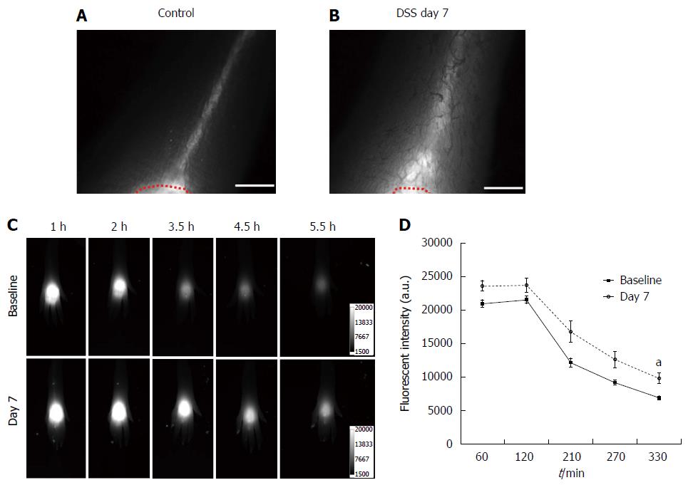
Figure 3 Near-infrared fluorescence imaging after id injection of indocyanine green in the dorsal aspect of the hind paw.
NIR fluorescent images of the foot in control mice (A) and mice treated with dextran sulfate sodium (DSS) for 7 d (B) after id injection of 2 μL of indocyanine green (ICG) in the dorsal aspect of the foot. A red dotted line delineates the ICG injection area. Representative fluorescent images in the foot of a mouse 1, 2, 3.5, 4.5, and 5.5 h after id injection of 2 μL of Alexa680-BSA prior to (baseline), and 7 d after DSS treatment displayed the clearance of Alexa680-BSA from the depot over time (C). Quantification of the fluorescent intensities (D) remaining in the depot of Alexa680-BSA in the skin of mice treated with DSS for 7 d (n = 7; grey open circle) showed higher fluorescent intensity over time as compared to baseline (filled black square), which was statistically significant at 5.5 h (P = 0.01) in comparison to control mice. aP < 0.05. Scale bar = 1 mm.
- Citation: Agollah GD, Wu G, Peng HL, Kwon S. Dextran sulfate sodium-induced acute colitis impairs dermal lymphatic function in mice. World J Gastroenterol 2015; 21(45): 12767-12777
- URL: https://www.wjgnet.com/1007-9327/full/v21/i45/12767.htm
- DOI: https://dx.doi.org/10.3748/wjg.v21.i45.12767


