Copyright
©The Author(s) 2015.
World J Gastroenterol. Nov 28, 2015; 21(44): 12713-12721
Published online Nov 28, 2015. doi: 10.3748/wjg.v21.i44.12713
Published online Nov 28, 2015. doi: 10.3748/wjg.v21.i44.12713
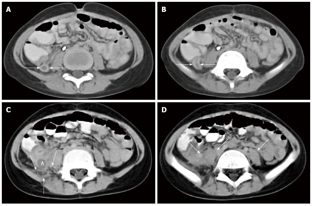
Figure 1 Computed tomography images.
Computed tomography images of abdomen using IV non-ionic contrast and standard oral contrast demonstrates dilated appendiceal ostium and thickened appendiceal wall in cross-section (arrows in A); dilated lumen and thickened wall of vermiform appendix in longitudinal section (arrows in B); periappendiceal fat stranding (arrows in C) and an enlarged right psoas muscle with indistinct margins from local extension of the appendiceal inflammation (enlarged right psoas muscle with indistinct margins identified in D by 2 arrows, as compared to normal-sized left psoas muscle with distinct margins identified by 1 arrow). The appendix measures approximately 11 mm in diameter from outer wall to outer wall. All these findings are consistent with acute appendicitis. There is no evident appendicolith or typhlitis.
-
Citation: Gjeorgjievski M, Amin MB, Cappell MS. Characteristic clinical features of
Aspergillus appendicitis: Case report and literature review. World J Gastroenterol 2015; 21(44): 12713-12721 - URL: https://www.wjgnet.com/1007-9327/full/v21/i44/12713.htm
- DOI: https://dx.doi.org/10.3748/wjg.v21.i44.12713


