Copyright
©The Author(s) 2015.
World J Gastroenterol. Nov 28, 2015; 21(44): 12544-12557
Published online Nov 28, 2015. doi: 10.3748/wjg.v21.i44.12544
Published online Nov 28, 2015. doi: 10.3748/wjg.v21.i44.12544
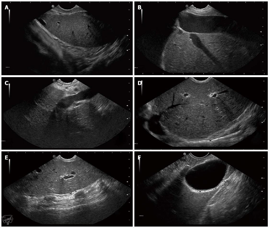
Figure 3 Endoscopic ultrasound images of the hepatic structures with the tip of the linear echoendoscope at different positions.
A: Endoscopic ultrasound (EUS) image of the left liver lobe with the diaphragm. The image is obtained from the cardia region; B: EUS image of the left liver lobe with the inferior vena cava and a hepatic vein; C: EUS image of the liver at the portal ligament region showing from the transducer, the hepatic artery, the portal vein and a short segment of the common bile duct. The transducer is located in the stomach; D: EUS image of the liver looking over the hepatic dome; E: EUS image of the right hepatic lobe. Note the shadows from the ribs at the anterior abdominal wall; F: EUS image of the liver with the gall bladder. The transducer is located in the first part of the duodenum.
- Citation: Srinivasan I, Tang SJ, Vilmann AS, Menachery J, Vilmann P. Hepatic applications of endoscopic ultrasound: Current status and future directions. World J Gastroenterol 2015; 21(44): 12544-12557
- URL: https://www.wjgnet.com/1007-9327/full/v21/i44/12544.htm
- DOI: https://dx.doi.org/10.3748/wjg.v21.i44.12544


