Copyright
©The Author(s) 2015.
World J Gastroenterol. Nov 21, 2015; 21(43): 12439-12447
Published online Nov 21, 2015. doi: 10.3748/wjg.v21.i43.12439
Published online Nov 21, 2015. doi: 10.3748/wjg.v21.i43.12439
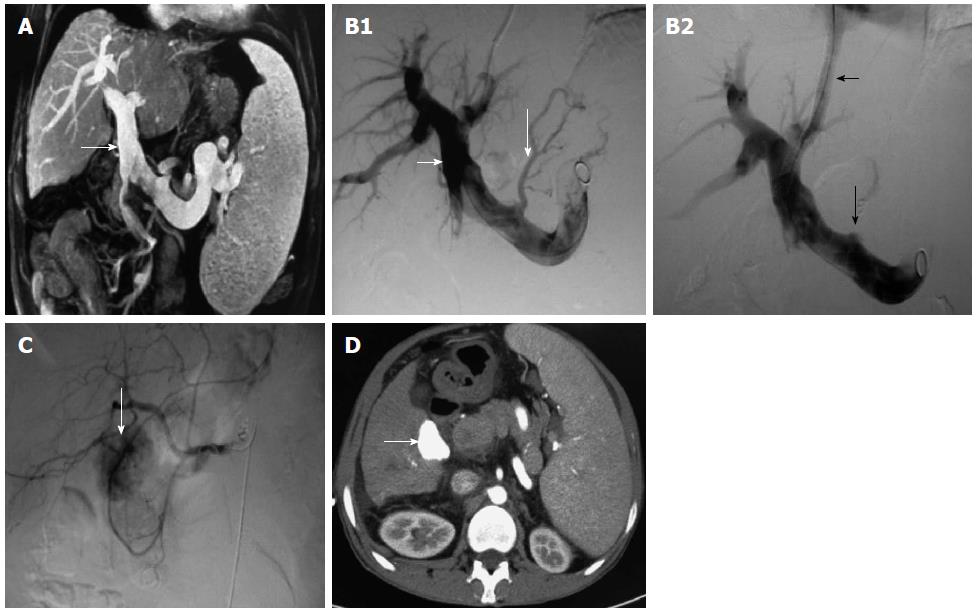
Figure 1 Patient with hepatitis C cirrhosis associated with esophageal gastric-fundus variceal bleeding was diagnosed with hepatocellular carcinoma 37 mo after transjugular intrahepatic portosystemic shunt.
We treated HCC with TACE. A: Obvious portal vein dilation (white arrow) revealed by magnetic resonance cholangiopancreatography; B1: Obvious portal vein dilation (short white arrow) and gastric coronary vein varicosis (long white arrow); B2: Gastric coronary vein embolism (long white arrow) and distributary channel (short white arrow); C: Tumor lesion with rich blood supply in the right hepatic lobe 37 mo after TIPS by hepatic angiography (white arrow); D: Iodine oil deposited in the lesion after TACE, shown by contrast-enhanced CT (white arrow). HCC: Hepatocellular carcinoma; TACE: Transarterial chemoembolization; TIPS: Transjugular intrahepatic portosystemic shunt; CT: Computed tomography.
- Citation: Qiu B, Zhao MF, Yue ZD, Zhao HW, Wang L, Fan ZH, He FL, Dai S, Yao JN, Liu FQ. Combined transjugular intrahepatic portosystemic shunt and other interventions for hepatocellular carcinoma with portal hypertension. World J Gastroenterol 2015; 21(43): 12439-12447
- URL: https://www.wjgnet.com/1007-9327/full/v21/i43/12439.htm
- DOI: https://dx.doi.org/10.3748/wjg.v21.i43.12439


