Copyright
©The Author(s) 2015.
World J Gastroenterol. Nov 21, 2015; 21(43): 12392-12402
Published online Nov 21, 2015. doi: 10.3748/wjg.v21.i43.12392
Published online Nov 21, 2015. doi: 10.3748/wjg.v21.i43.12392
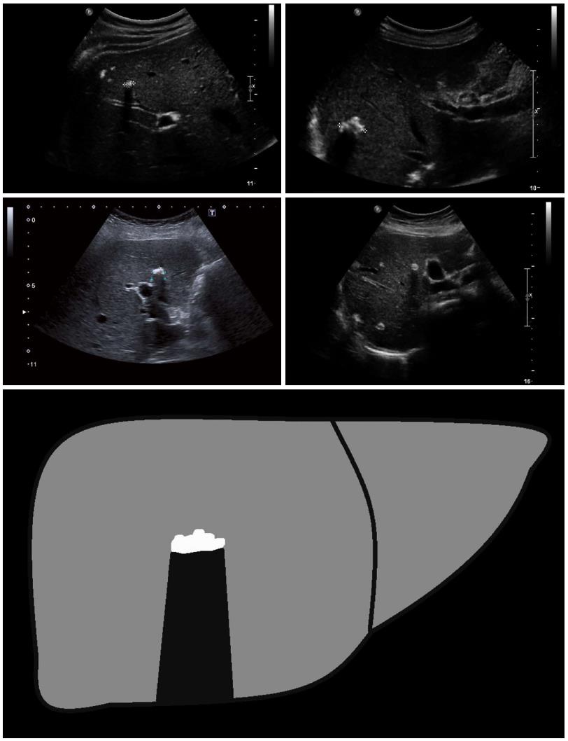
Figure 4 Ossification: The ossification pattern presents with solitary or grouped, mostly sharply delineated lesions with dorsal acoustic shadow.
In terms of their differential diagnosis, these alveolar echinococcosis (AE) lesions are often difficult to distinguish from inflammatory or hyperechoic metastases of various carcinomas. Very large ossification-type AE lesions represent a rarity. Both uni- and multifocal involvement is possible.
- Citation: Kratzer W, Gruener B, Kaltenbach TE, Ansari-Bitzenberger S, Kern P, Fuchs M, Mason RA, Barth TF, Haenle MM, Hillenbrand A, Oeztuerk S, Graeter T. Proposal of an ultrasonographic classification for hepatic alveolar echinococcosis: Echinococcosis multilocularis Ulm classification-ultrasound. World J Gastroenterol 2015; 21(43): 12392-12402
- URL: https://www.wjgnet.com/1007-9327/full/v21/i43/12392.htm
- DOI: https://dx.doi.org/10.3748/wjg.v21.i43.12392


