Copyright
©The Author(s) 2015.
World J Gastroenterol. Nov 7, 2015; 21(41): 11842-11853
Published online Nov 7, 2015. doi: 10.3748/wjg.v21.i41.11842
Published online Nov 7, 2015. doi: 10.3748/wjg.v21.i41.11842
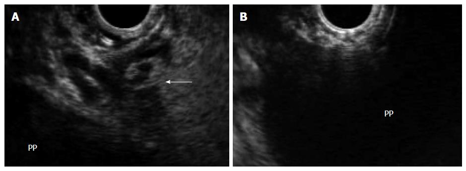
Figure 3 In the same patient, endoscopic ultrasound permits visualization of the pancreatic pseudocyst, assessment of wall maturity, determination of distance between the collection and the luminal wall, identification of intervening vessels or collaterals (arrow) (A), and selection of the optimal puncture site (B).
PP: Pancreatic pseudocyst.
- Citation: Vilmann AS, Menachery J, Tang SJ, Srinivasan I, Vilmann P. Endosonography guided management of pancreatic fluid collections. World J Gastroenterol 2015; 21(41): 11842-11853
- URL: https://www.wjgnet.com/1007-9327/full/v21/i41/11842.htm
- DOI: https://dx.doi.org/10.3748/wjg.v21.i41.11842


