Copyright
©The Author(s) 2015.
World J Gastroenterol. Nov 7, 2015; 21(41): 11832-11841
Published online Nov 7, 2015. doi: 10.3748/wjg.v21.i41.11832
Published online Nov 7, 2015. doi: 10.3748/wjg.v21.i41.11832
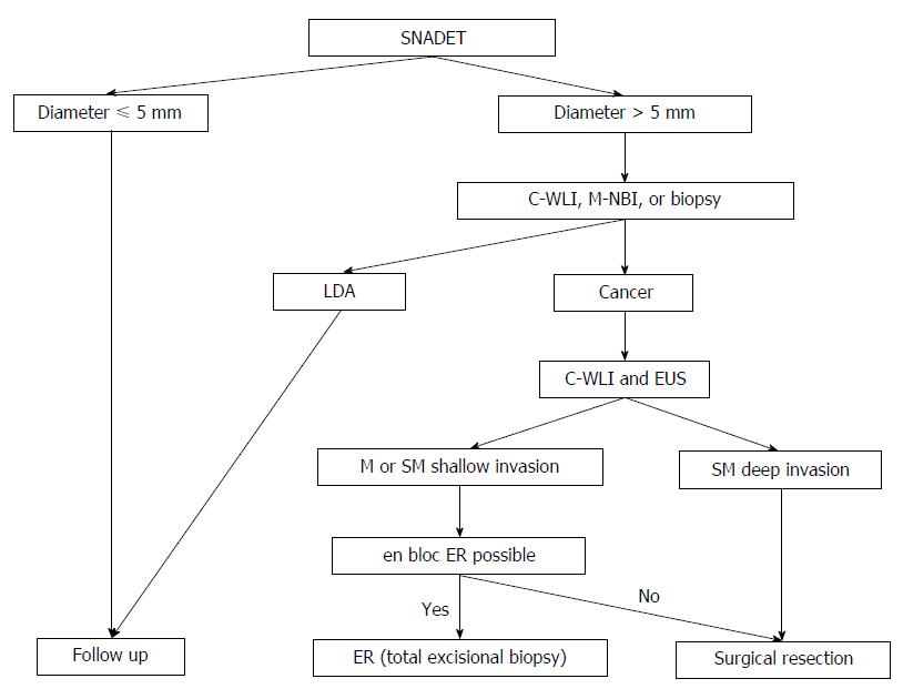
Figure 8 Suggested algorithm for the management of superficial non-ampullary duodenal epithelial tumor according to depth of invasion; tumor size; endoscopic findings, including magnifying endoscopy; and biopsy results.
Endoscopic features of cancer are a red color in the tumor; a nodular, rough surface on conventional white light imaging; a marginal type of milk-white mucosa; an unclassified vascular pattern; a frequency of ill-defined mucosal pattern; and a population of mixed-type lesions with multiple surface patterns on magnifying endoscopy with narrow-band imaging. Endoscopic features of submucosal carcinoma are ulceration and a 0-I or 0-IIa + IIc macroscopic type with a red color. SNADET: Superficial non-ampullary duodenal epithelial tumor; C-WLI: Conventional white-light imaging; M-NBI: Magnifying endoscopy with narrow-band imaging; LDA: Low-grade adenoma; EUS: Endoscopic ultrasonography; ER: Endoscopic resection.
- Citation: Tsuji S, Doyama H, Tsuji K, Tsuyama S, Tominaga K, Yoshida N, Takemura K, Yamada S, Niwa H, Katayanagi K, Kurumaya H, Okada T. Preoperative endoscopic diagnosis of superficial non-ampullary duodenal epithelial tumors, including magnifying endoscopy. World J Gastroenterol 2015; 21(41): 11832-11841
- URL: https://www.wjgnet.com/1007-9327/full/v21/i41/11832.htm
- DOI: https://dx.doi.org/10.3748/wjg.v21.i41.11832


