Copyright
©The Author(s) 2015.
World J Gastroenterol. Nov 7, 2015; 21(41): 11832-11841
Published online Nov 7, 2015. doi: 10.3748/wjg.v21.i41.11832
Published online Nov 7, 2015. doi: 10.3748/wjg.v21.i41.11832
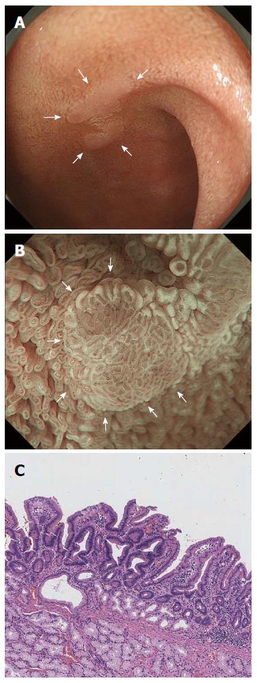
Figure 5 Magnifying endoscopy with narrow-band imaging imaging of a duodenal adenoma.
A: Endoscopic findings using conventional endoscopy with white light imaging. A pale, slightly elevated lesion (10 mm in diameter, arrow) is observed in the proximal duodenum; B: Endoscopic findings using magnifying endoscopy with narrow-band imaging (M-NBI). A demarcation line (DL, arrows) separates changes in the mucosal microsurface (MS) structure from the surrounding normal mucosa. Vessel plus surface (VS) classifications: V, Because of the white opaque substance (WOS), the morphology of the subepithelial microvessels cannot be observed, making this an absent microvascular (MV) pattern; S, The WOS has a regular reticular pattern with a symmetrical distribution and regular arrangement. Thus, this lesion is graded as a regular MS pattern using WOS as a marker for the MS pattern. The VS classification of this lesion was absent MV pattern and regular MS pattern (WOS+) with a DL. Therefore, the M-NBI diagnosis was benign; C: The final histological diagnosis was of a low-grade adenoma.
- Citation: Tsuji S, Doyama H, Tsuji K, Tsuyama S, Tominaga K, Yoshida N, Takemura K, Yamada S, Niwa H, Katayanagi K, Kurumaya H, Okada T. Preoperative endoscopic diagnosis of superficial non-ampullary duodenal epithelial tumors, including magnifying endoscopy. World J Gastroenterol 2015; 21(41): 11832-11841
- URL: https://www.wjgnet.com/1007-9327/full/v21/i41/11832.htm
- DOI: https://dx.doi.org/10.3748/wjg.v21.i41.11832


