Copyright
©The Author(s) 2015.
World J Gastroenterol. Nov 7, 2015; 21(41): 11832-11841
Published online Nov 7, 2015. doi: 10.3748/wjg.v21.i41.11832
Published online Nov 7, 2015. doi: 10.3748/wjg.v21.i41.11832
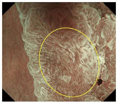
Figure 4 Duodenal adenocarcinoma imaged with magnifying endoscopy with narrow-band imaging.
Because of uneven distribution of white opaque substance (WOS) on magnifying endoscopy with narrow-band imaging (M-NBI), this lesion displays multiple microsurface patterns as mixed-type (yellow circle).
- Citation: Tsuji S, Doyama H, Tsuji K, Tsuyama S, Tominaga K, Yoshida N, Takemura K, Yamada S, Niwa H, Katayanagi K, Kurumaya H, Okada T. Preoperative endoscopic diagnosis of superficial non-ampullary duodenal epithelial tumors, including magnifying endoscopy. World J Gastroenterol 2015; 21(41): 11832-11841
- URL: https://www.wjgnet.com/1007-9327/full/v21/i41/11832.htm
- DOI: https://dx.doi.org/10.3748/wjg.v21.i41.11832


