Copyright
©The Author(s) 2015.
World J Gastroenterol. Nov 7, 2015; 21(41): 11502-11521
Published online Nov 7, 2015. doi: 10.3748/wjg.v21.i41.11502
Published online Nov 7, 2015. doi: 10.3748/wjg.v21.i41.11502
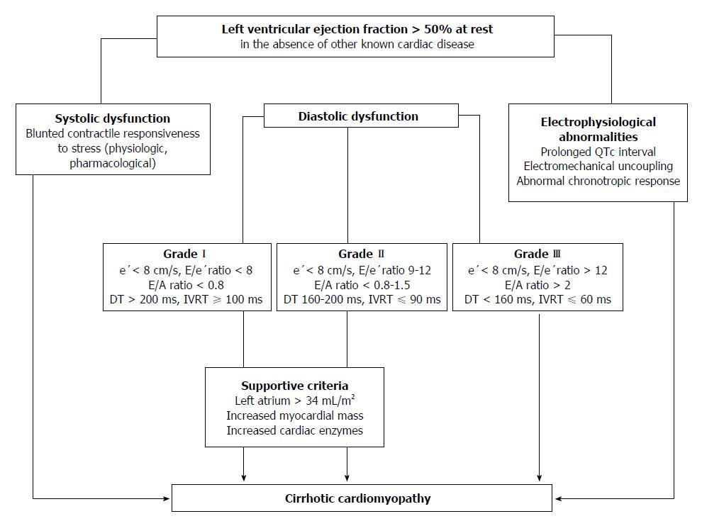
Figure 3 Algorithm for the diagnosis of cirrhotic cardiomyopathy.
Three ways to diagnose CCM with normal EF at rest have been displayed: (1) Systolic function. Patients have documented blunted responsiveness to volume and postural challenge, exercise, or pharmacological infusion but the manoeuvres that can bring systolic dysfunction have not been standardized yet; (2) Diastolic function. The diagnosis of LVDD can be obtained by TDI (E/e′ > 15). If TDI yields an E/e′ ratio (8 < E/e′ < 15) other echocardiographic investigations such as blood flow Doppler of mitral valve or pulmonary veins, LV mass index or left atrial volume index are required for diagnostic of LVDD; (3) Electrophysiological abnormalities. a, the prolongation of the electrocardiographic corrected QT interval is common in cirrhosis; b, electromechanical uncoupling is a dyssynchrony between electrical and mechanical systole. The electrical systole is longer in patients with cirrhosis; c, chronotropic incompetence is the inability of the heart to proportionally increase HR in response to stimuli (exercise, tilting, paracentesis, infections and pharmacological agents). CCM: Cirrhotic cardiomyopathy; EF: Ejection fraction; LVDD: Left ventricular diastolic dysfunction; TDI: Tissue Doppler imaging; e’: Peak early diastolic velocity at the basal part of the septal and lateral corner of the mitral annulus; E/e’ratio: Peak E-wave transmitral/early diastolic mitral annular velocity; E/A ratio: Early diastolic mitral inflow velocity/late diastolic velocity; DT: Deceleration time; IVRT: Isovolumic relaxation time.
- Citation: Ruiz-del-Árbol L, Serradilla R. Cirrhotic cardiomyopathy. World J Gastroenterol 2015; 21(41): 11502-11521
- URL: https://www.wjgnet.com/1007-9327/full/v21/i41/11502.htm
- DOI: https://dx.doi.org/10.3748/wjg.v21.i41.11502


