Copyright
©The Author(s) 2015.
World J Gastroenterol. Oct 28, 2015; 21(40): 11411-11427
Published online Oct 28, 2015. doi: 10.3748/wjg.v21.i40.11411
Published online Oct 28, 2015. doi: 10.3748/wjg.v21.i40.11411
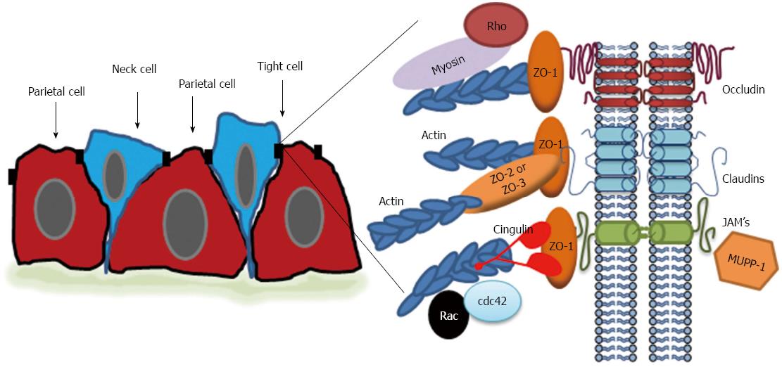
Figure 3 Schematic representation of the tight junction in gastric epithelial cells.
In the neck region of gastric glands, neck and parietal cells are oddly shaped but make tight junctions at the apical border between cells. Expanded diagram: Tight junctions in the stomach have classical components consisting of transmembrane proteins including occludin, claudins, and JAM proteins; peripheral scaffolding proteins like zonula occludens (ZO)-1, 2, and 3; linker proteins to the actin cytoskeleton like non-muscle myosin and cingulin; and signaling molecules like Rho, Rac, and cdc42. Actin filaments are also prominent.
-
Citation: Caron TJ, Scott KE, Fox JG, Hagen SJ. Tight junction disruption:
Helicobacter pylori and dysregulation of the gastric mucosal barrier. World J Gastroenterol 2015; 21(40): 11411-11427 - URL: https://www.wjgnet.com/1007-9327/full/v21/i40/11411.htm
- DOI: https://dx.doi.org/10.3748/wjg.v21.i40.11411


