Copyright
©The Author(s) 2015.
World J Gastroenterol. Oct 28, 2015; 21(40): 11396-11410
Published online Oct 28, 2015. doi: 10.3748/wjg.v21.i40.11396
Published online Oct 28, 2015. doi: 10.3748/wjg.v21.i40.11396
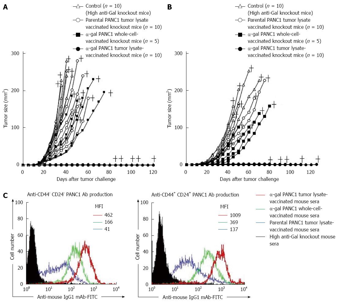
Figure 8 In vivo tumor growth in adoptively transferred non-obese diabetic/severe combined immunodeficiency mice challenged with either live PANC1 cells or live CD44+CD24+ PANC1 cells, and production of antibodies against differentiated cancer cells and cancer stem cells.
A: We monitored tumor growth in splenocyte-transferred mice. No tumors were noted in the α-gal tumor lysate-vaccinated mice. No significant differences in the time to appearance of a palpable tumor after tumor challenge were observed in the untreated control group and parental tumor lysate group (untreated: 10.6 ± 2.5 d; parental tumor lysate: 11.9 ± 2.1 d). In contrast, the development of tumors in the α-gal whole cell vaccination group was significantly delayed compared with the untreated and parental tumor lysate groups (α-gal whole-cell: 16.0 ± 2.8 d, P = 0.018 vs control; P = 0.004 vs parental tumor lysate). In the untreated control group, the maximum tumor size was 100 mm2 within 29 to 34 d (mean: 31.4 ± 2.1 d). In comparison, tumor growth to a similar size was markedly delayed in both the parental tumor lysate group (40.3 ± 6.9 d, P = 0.007 vs control) and α-gal whole-cell group (45.6 ± 8.3 d, P = 0.0013 vs control). +; death; B: The tumorigenesis of pancreatic CSCs was completely prevented in all α-gal tumor lysate-vaccinated mice. With the exception of the α-gal tumor lysate group, no significant differences were seen in the time to appearance of palpable tumors after tumor challenge among the groups (untreated: 13.1 ± 3.3 d; parental tumor lysate: 14.4 ± 3.4 d; α-gal whole-cell: 17.0 ± 3.8 d). The tumor size reached 100 mm2 in 40.6 ± 1.8 and 48.0 ± 4.4 d in the untreated and parental tumor lysate groups, respectively. However, tumor growth to a similar size was significantly delayed in the α-gal whole-cell group (60.5 ± 7.9 d; P < 0.001 vs control; P = 0.033 vs parental tumor lysate). +; death; C: Production of either anti-CD44-CD24- PANC1 (i.e., differentiated pancreatic cancer cells) antibodies or anti-CD44+CD24+ PANC1 (i.e., pancreatic cancer stem cells) antibodies in sera of vaccinated knockout mice assessed with flow cytometry. Closed histogram; stained cells with sera from non-vaccinated knockout mice, open histogram; stained cells with sera from vaccinated knockout mice. MFI: Mean fluorescence intensity.
- Citation: Tanemura M, Miyoshi E, Nagano H, Eguchi H, Matsunami K, Taniyama K, Hatanaka N, Akamatsu H, Mori M, Doki Y. Cancer immunotherapy for pancreatic cancer utilizing α-gal epitope/natural anti-Gal antibody reaction. World J Gastroenterol 2015; 21(40): 11396-11410
- URL: https://www.wjgnet.com/1007-9327/full/v21/i40/11396.htm
- DOI: https://dx.doi.org/10.3748/wjg.v21.i40.11396


