Copyright
©The Author(s) 2015.
World J Gastroenterol. Jan 28, 2015; 21(4): 1349-1356
Published online Jan 28, 2015. doi: 10.3748/wjg.v21.i4.1349
Published online Jan 28, 2015. doi: 10.3748/wjg.v21.i4.1349
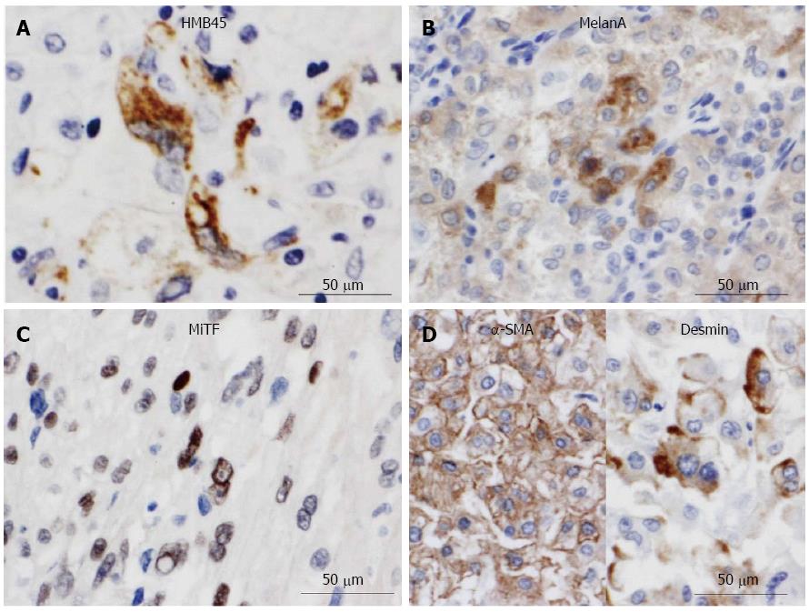
Figure 4 Immunohistochemical examination of malignant perivascular epithelioid cell tumor of the stomach.
The highly atypical epithelioid cells were specifically positive for melanocytic markers, such as HMB45 (A), Melan A (B), and microphthalmia transcription factor (MiTF) (C), and muscle markers, such as α-smooth muscle actin (SMA) markers (D) or desmin (D). Bars = 50 μm.
- Citation: Yamada S, Nabeshima A, Noguchi H, Nawata A, Nishii H, Guo X, Wang KY, Hisaoka M, Nakayama T. Coincidence between malignant perivascular epithelioid cell tumor arising in the gastric serosa and lung adenocarcinoma. World J Gastroenterol 2015; 21(4): 1349-1356
- URL: https://www.wjgnet.com/1007-9327/full/v21/i4/1349.htm
- DOI: https://dx.doi.org/10.3748/wjg.v21.i4.1349


