Copyright
©The Author(s) 2015.
World J Gastroenterol. Jan 28, 2015; 21(4): 1349-1356
Published online Jan 28, 2015. doi: 10.3748/wjg.v21.i4.1349
Published online Jan 28, 2015. doi: 10.3748/wjg.v21.i4.1349
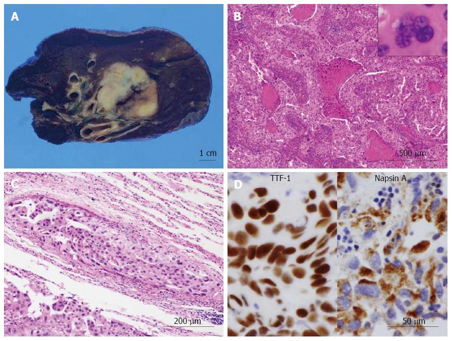
Figure 2 Gross, histological and immunohistochemical findings of poorly differentiated adenocarcinoma of the lung.
A: On gross examination, the cut surface showed a solid firm and lobulated mass, measuring 32 mm × 30 mm × 29 mm, which looked from gray-whitish to yellow-whitish in color, accompanied with focal necrosis and hemorrhage. The background had no remarkable change. Bar = 1 cm; B: Low to medium power view exhibited a proliferation of medium-sized to large atypical epithelial cells having hyperchromatic pleomorphic nuclei and abundant eosinophilic cytoplasm, predominantly arranged in an acinar or solid fashion with frequent necrotic foci (HE stains). Multi-nucleated giant tumor cells were readily encountered (inset). Bar = 500 μm; C: The tumor nests peripherally involved the vascular vessel (HE stains). Bar = 200 μm; D: In immunohistochemistry, these adenocarcinoma cells were specifically positive for thyroid transcription factor 1 (TTF-1) (left) and Napsin A (right), in nuclear and intracytoplasmic pattern, respectively. Bar = 50 μm.
- Citation: Yamada S, Nabeshima A, Noguchi H, Nawata A, Nishii H, Guo X, Wang KY, Hisaoka M, Nakayama T. Coincidence between malignant perivascular epithelioid cell tumor arising in the gastric serosa and lung adenocarcinoma. World J Gastroenterol 2015; 21(4): 1349-1356
- URL: https://www.wjgnet.com/1007-9327/full/v21/i4/1349.htm
- DOI: https://dx.doi.org/10.3748/wjg.v21.i4.1349


