Copyright
©The Author(s) 2015.
World J Gastroenterol. Jan 28, 2015; 21(4): 1349-1356
Published online Jan 28, 2015. doi: 10.3748/wjg.v21.i4.1349
Published online Jan 28, 2015. doi: 10.3748/wjg.v21.i4.1349
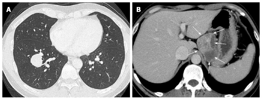
Figure 1 Findings of chest and abdominal computed tomography scan at surgery.
A: A chest computed tomography (CT) scan showed a relatively well-demarcated nodule, measuring approximately 30 mm × 30 mm, in the right lower lobe, S9; B: An abdominal CT scan showed a relatively well-defined huge mass with heterogeneously enhancement (arrows), measuring approximately 70 mm × 60 mm, attached to the gastric wall and separated from the left kidney and adrenal gland.
- Citation: Yamada S, Nabeshima A, Noguchi H, Nawata A, Nishii H, Guo X, Wang KY, Hisaoka M, Nakayama T. Coincidence between malignant perivascular epithelioid cell tumor arising in the gastric serosa and lung adenocarcinoma. World J Gastroenterol 2015; 21(4): 1349-1356
- URL: https://www.wjgnet.com/1007-9327/full/v21/i4/1349.htm
- DOI: https://dx.doi.org/10.3748/wjg.v21.i4.1349


