Copyright
©The Author(s) 2015.
World J Gastroenterol. Jan 28, 2015; 21(4): 1344-1348
Published online Jan 28, 2015. doi: 10.3748/wjg.v21.i4.1344
Published online Jan 28, 2015. doi: 10.3748/wjg.v21.i4.1344
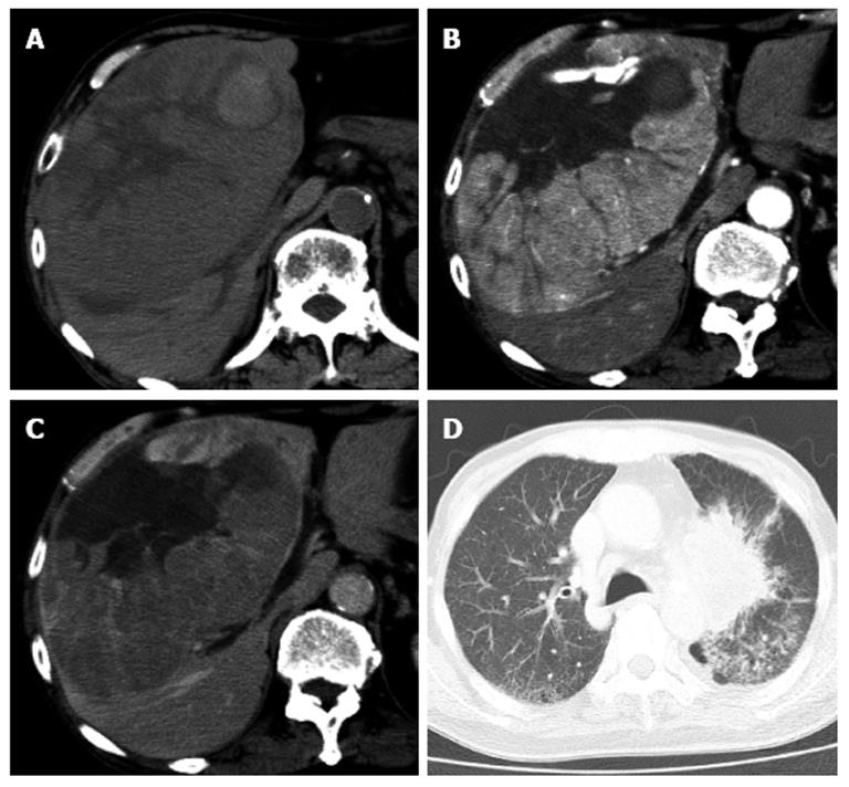
Figure 1 Contrast enhanced computed tomography image of the abdomen shows a huge tumor that occupies the right hepatic lobe, with enhancement in a viable lesion.
A: Non-contrast phase; B: Hepatic arterial phase; C: Portal vein phase; D: Computed tomography image of the chest shows a primary lung cancer in the hilar side of the left lung.
- Citation: Hatamaru K, Azuma S, Akamatsu T, Seta T, Urai S, Uenoyama Y, Yamashita Y, Ono K. Pulmonary embolism after arterial chemoembolization for hepatocellular carcinoma: An autopsy case report. World J Gastroenterol 2015; 21(4): 1344-1348
- URL: https://www.wjgnet.com/1007-9327/full/v21/i4/1344.htm
- DOI: https://dx.doi.org/10.3748/wjg.v21.i4.1344


