Copyright
©The Author(s) 2015.
World J Gastroenterol. Jan 28, 2015; 21(4): 1334-1343
Published online Jan 28, 2015. doi: 10.3748/wjg.v21.i4.1334
Published online Jan 28, 2015. doi: 10.3748/wjg.v21.i4.1334
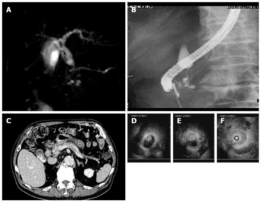
Figure 6 Imaging findings of Case 5.
A, B: Stenosis in the lower extrahepatic bile duct and a slight dilation of the main pancreatic duct on endoscopic retrograde cholangiopancreatography (A) and magnetic resonance cholangiopancreatography (B); C: Normal size of the pancreas on abdominal computed tomography; D (at the hilar hepatic lesion); E (at the bifurcation of cystic duct); F (at the intrapancreatic lesion): Bile duct wall thickening with smooth inner and outer margin in areas with stenosis (F) and without (D, E) on intraductal ultrasonography.
- Citation: Nakazawa T, Ikeda Y, Kawaguchi Y, Kitagawa H, Takada H, Takeda Y, Makino I, Makino N, Naitoh I, Tanaka A. Isolated intrapancreatic IgG4-related sclerosing cholangitis. World J Gastroenterol 2015; 21(4): 1334-1343
- URL: https://www.wjgnet.com/1007-9327/full/v21/i4/1334.htm
- DOI: https://dx.doi.org/10.3748/wjg.v21.i4.1334


