Copyright
©The Author(s) 2015.
World J Gastroenterol. Jan 28, 2015; 21(4): 1329-1333
Published online Jan 28, 2015. doi: 10.3748/wjg.v21.i4.1329
Published online Jan 28, 2015. doi: 10.3748/wjg.v21.i4.1329
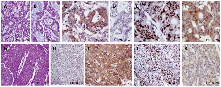
Figure 1 Cecum mixed adenoneuroendocrine carcinoma consists of proliferation of two components.
First component is represented by glandular structures with mucinous foci (A-B) and displays positivity for keratin 20 (C) without immunoreactivity for synaptophysin (D), with a high Ki67 index (E) and cytoplasmic expression of maspin (F); The second component is represented by solid tumor islands (G) that are negative for keratin 20 (H) and are immunoreactive for synaptophysin (I), with a lower Ki67 index (J) and slightly cytoplasmic expression of maspin (K).
- Citation: Gurzu S, Kadar Z, Bara T, Bara TJ, Tamasi A, Azamfirei L, Jung I. Mixed adenoneuroendocrine carcinoma of gastrointestinal tract: Report of two cases. World J Gastroenterol 2015; 21(4): 1329-1333
- URL: https://www.wjgnet.com/1007-9327/full/v21/i4/1329.htm
- DOI: https://dx.doi.org/10.3748/wjg.v21.i4.1329


