Copyright
©The Author(s) 2015.
World J Gastroenterol. Jan 28, 2015; 21(4): 1324-1328
Published online Jan 28, 2015. doi: 10.3748/wjg.v21.i4.1324
Published online Jan 28, 2015. doi: 10.3748/wjg.v21.i4.1324
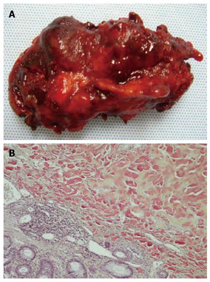
Figure 3 Five cm amyloidoma specimen excised from the rectum 2 cm from the dentate line (A) and photomicrograph (x 40) demonstrating amyloid deposition with Congo red staining in rectal amyloidoma (B).
Amyloid protein is seen as acellular, homogeneous, and eosinphilic material deposited uniformly throughout the mass.
- Citation: Sharma R, George VV. Transanal endoscopic microsurgery: The first attempt in treatment of rectal amyloidoma. World J Gastroenterol 2015; 21(4): 1324-1328
- URL: https://www.wjgnet.com/1007-9327/full/v21/i4/1324.htm
- DOI: https://dx.doi.org/10.3748/wjg.v21.i4.1324


