Copyright
©The Author(s) 2015.
World J Gastroenterol. Jan 28, 2015; 21(4): 1324-1328
Published online Jan 28, 2015. doi: 10.3748/wjg.v21.i4.1324
Published online Jan 28, 2015. doi: 10.3748/wjg.v21.i4.1324
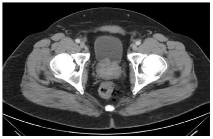
Figure 1 Computed tomography of the pelvis demonstrates a thickening of the left lower rectal wall with adjacent free gas.
No pathologically enlarged lymphadenopathy was found. Arrow mark the extraluminal air or free air in the rectum.
- Citation: Sharma R, George VV. Transanal endoscopic microsurgery: The first attempt in treatment of rectal amyloidoma. World J Gastroenterol 2015; 21(4): 1324-1328
- URL: https://www.wjgnet.com/1007-9327/full/v21/i4/1324.htm
- DOI: https://dx.doi.org/10.3748/wjg.v21.i4.1324


