Copyright
©The Author(s) 2015.
World J Gastroenterol. Jan 28, 2015; 21(4): 1091-1098
Published online Jan 28, 2015. doi: 10.3748/wjg.v21.i4.1091
Published online Jan 28, 2015. doi: 10.3748/wjg.v21.i4.1091
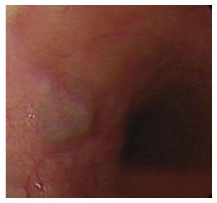
Figure 9 Hemangioma.
On endoscopy, esophageal hemangioma appears cystic and bluish-red and can be pressed with biopsy forceps.
- Citation: Tsai SJ, Lin CC, Chang CW, Hung CY, Shieh TY, Wang HY, Shih SC, Chen MJ. Benign esophageal lesions: Endoscopic and pathologic features. World J Gastroenterol 2015; 21(4): 1091-1098
- URL: https://www.wjgnet.com/1007-9327/full/v21/i4/1091.htm
- DOI: https://dx.doi.org/10.3748/wjg.v21.i4.1091


