Copyright
©The Author(s) 2015.
World J Gastroenterol. Jan 28, 2015; 21(4): 1091-1098
Published online Jan 28, 2015. doi: 10.3748/wjg.v21.i4.1091
Published online Jan 28, 2015. doi: 10.3748/wjg.v21.i4.1091
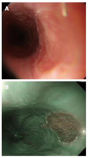
Figure 1 Heterotopic gastric mucosa.
A: Heterotopic gastric mucosa appears salmon-colored under conventional endoscopy and is recognized as flat or slightly elevated; B: Narrow band imaging facilitates mucosal surface evaluation of heterotopic gastric mucosa by adjusting reflected light to enhance the contrast between the esophageal mucosa and the gastric mucosa and may improve the diagnosis of heterotopic gastric mucosa.
- Citation: Tsai SJ, Lin CC, Chang CW, Hung CY, Shieh TY, Wang HY, Shih SC, Chen MJ. Benign esophageal lesions: Endoscopic and pathologic features. World J Gastroenterol 2015; 21(4): 1091-1098
- URL: https://www.wjgnet.com/1007-9327/full/v21/i4/1091.htm
- DOI: https://dx.doi.org/10.3748/wjg.v21.i4.1091


