Copyright
©The Author(s) 2015.
World J Gastroenterol. Oct 21, 2015; 21(39): 11199-11204
Published online Oct 21, 2015. doi: 10.3748/wjg.v21.i39.11199
Published online Oct 21, 2015. doi: 10.3748/wjg.v21.i39.11199
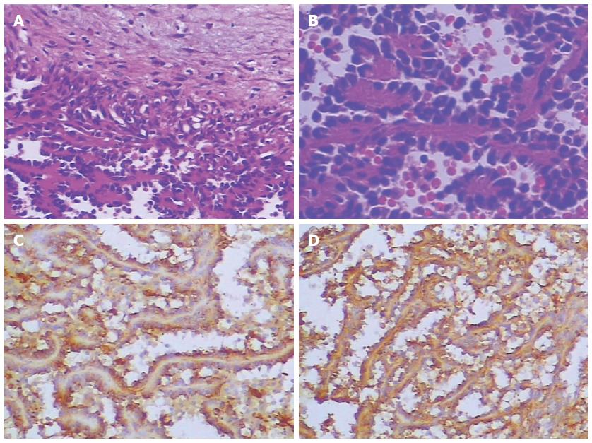
Figure 3 Histological chrematistics of angiosarcoma.
A: Histological analysis showing spindled vascular proliferation and area of necrosis (HE staining, × 200); B: Histological analysis showing atypical spindle cells with few mitotic figures (HE staining, × 400); C: Immunohistochemical analysis showing the lesion stained positively for CD31 (× 400); D: Immunohistochemical analysis showing the lesion stained positively for F8 (× 400).
- Citation: Chen F, Jin HF, Fan YH, Cai LJ, Zhang ZY, Lv B. Case report of primary splenic angiosarcoma with hepatic metastases. World J Gastroenterol 2015; 21(39): 11199-11204
- URL: https://www.wjgnet.com/1007-9327/full/v21/i39/11199.htm
- DOI: https://dx.doi.org/10.3748/wjg.v21.i39.11199


