Copyright
©The Author(s) 2015.
World J Gastroenterol. Oct 21, 2015; 21(39): 10936-10947
Published online Oct 21, 2015. doi: 10.3748/wjg.v21.i39.10936
Published online Oct 21, 2015. doi: 10.3748/wjg.v21.i39.10936
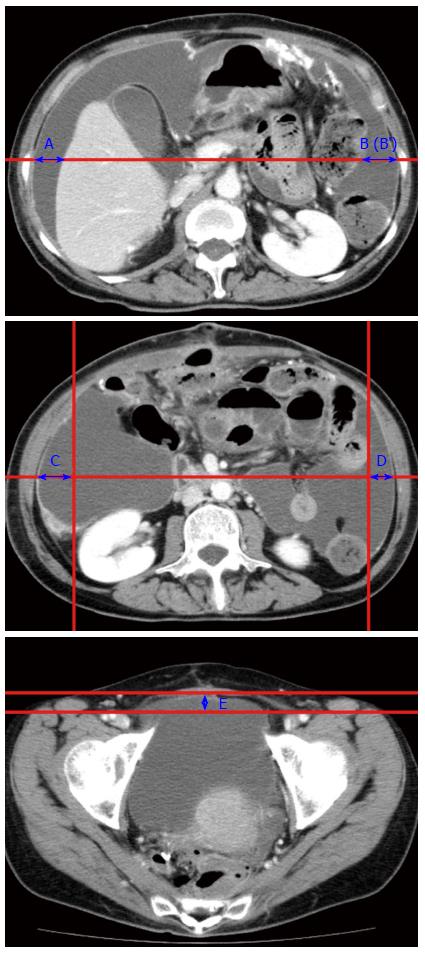
Figure 1 Five-point method to measure ascites volume.
Upper: Line between the bilateral antero-posterior mid-points of the abdominal wall is drawn at the plane of the root of the superior mesenteric artery. The distances between the inner surface of the right abdominal wall and the liver (A cm), and between the inner surface of the left abdominal wall and spleen (B cm) are obtained. When spleen is not observed on this plane, the distance between the left abdominal wall and margin to the ascites and internal organs are measured (B’ cm); Middle: The lower pole of the left kidney is observed on this plane. The sagittal line from the bilateral paracolic gutter, and between the bilateral antero-posterior midpoints of the abdominal wall is drawn. The distances C (cm) and D (cm) are thus obtained. Lower: A line between the anterior sides of the bilateral femoral artery is drawn. The distance between the inner surface of the abdomen (at the middle) and the line is obtained (E cm). The ascites volume is calculated by the equation of (A + B + C + D + E) × 200 (mL).
- Citation: Maeda H, Kobayashi M, Sakamoto J. Evaluation and treatment of malignant ascites secondary to gastric cancer. World J Gastroenterol 2015; 21(39): 10936-10947
- URL: https://www.wjgnet.com/1007-9327/full/v21/i39/10936.htm
- DOI: https://dx.doi.org/10.3748/wjg.v21.i39.10936


