Copyright
©The Author(s) 2015.
World J Gastroenterol. Oct 14, 2015; 21(38): 10926-10930
Published online Oct 14, 2015. doi: 10.3748/wjg.v21.i38.10926
Published online Oct 14, 2015. doi: 10.3748/wjg.v21.i38.10926
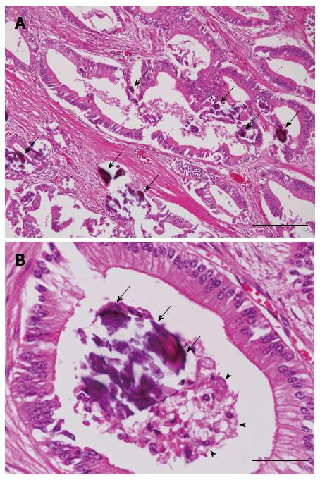
Figure 5 Histopathological findings (hematoxylin and eosin staining).
A: Microscopic examination reveals a mucus-secreting, gastric foveolar type adenocarcinoma with numerous fine calcifications (arrows) (bar = 200 μm); B: The calcified material (arrows) is located in the mucus (arrow head) in the tumor glands (bar = 50 μm).
- Citation: Inoko K, Tsuchikawa T, Noji T, Kurashima Y, Ebihara Y, Tamoto E, Nakamura T, Murakami S, Okamura K, Shichinohe T, Hirano S. Hilar cholangiocarcinoma with intratumoral calcification: A case report. World J Gastroenterol 2015; 21(38): 10926-10930
- URL: https://www.wjgnet.com/1007-9327/full/v21/i38/10926.htm
- DOI: https://dx.doi.org/10.3748/wjg.v21.i38.10926


