Copyright
©The Author(s) 2015.
World J Gastroenterol. Oct 14, 2015; 21(38): 10926-10930
Published online Oct 14, 2015. doi: 10.3748/wjg.v21.i38.10926
Published online Oct 14, 2015. doi: 10.3748/wjg.v21.i38.10926
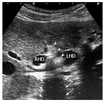
Figure 1 Finding of ultrasonography.
Transverse ultrasonography shows a highly echogenic mass (arrow head) with posterior acoustic shadowing (arrow) at the confluence of the right and left hepatic duct. RHD: Right hepatic duct; LHD: Left hepatic duct.
- Citation: Inoko K, Tsuchikawa T, Noji T, Kurashima Y, Ebihara Y, Tamoto E, Nakamura T, Murakami S, Okamura K, Shichinohe T, Hirano S. Hilar cholangiocarcinoma with intratumoral calcification: A case report. World J Gastroenterol 2015; 21(38): 10926-10930
- URL: https://www.wjgnet.com/1007-9327/full/v21/i38/10926.htm
- DOI: https://dx.doi.org/10.3748/wjg.v21.i38.10926


