Copyright
©The Author(s) 2015.
World J Gastroenterol. Oct 14, 2015; 21(38): 10840-10852
Published online Oct 14, 2015. doi: 10.3748/wjg.v21.i38.10840
Published online Oct 14, 2015. doi: 10.3748/wjg.v21.i38.10840
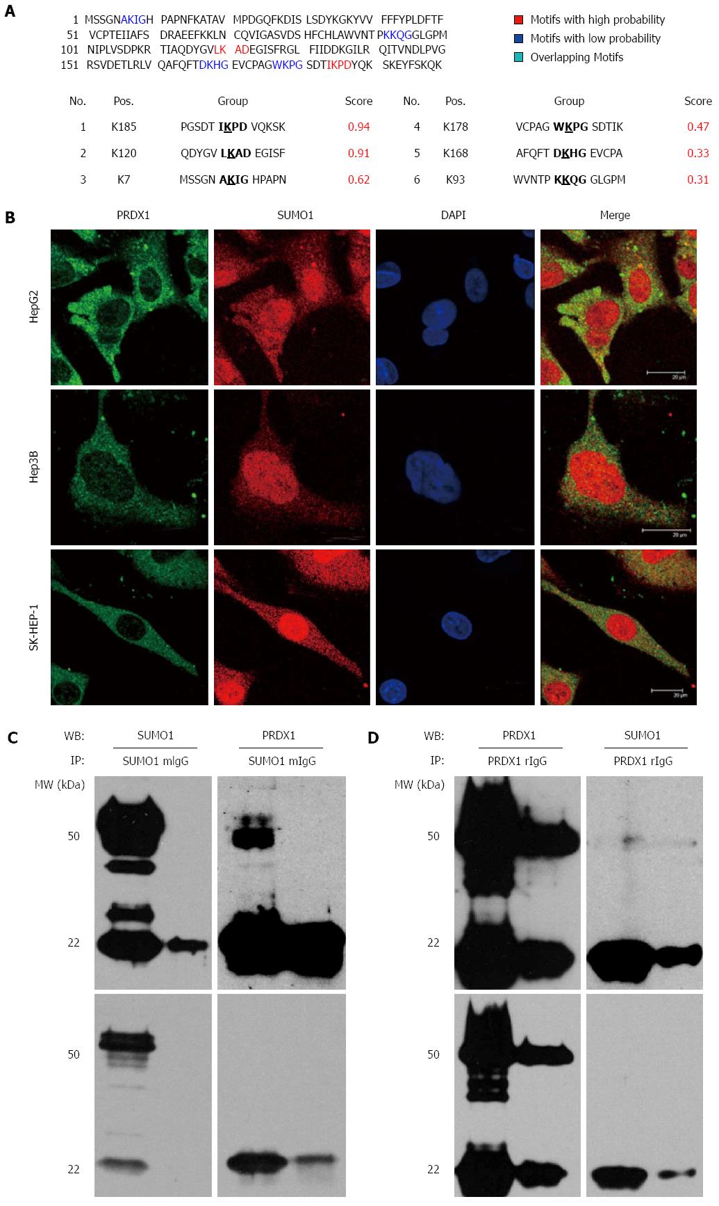
Figure 4 Peroxiredoxin 1 might be sumoylated in liver cancer cells.
A: The bioinformatic prediction of PRDX1 using SUMOplot tool; B: Immunofluorescence staining visualized under a confocal microscope illustrating the co-localization of PRDX1 and SUMO1 proteins in the cytoplasm of three liver cancer cells. C, D: Co-immunoprecipitation of PRDX1 with SUMO1 in HepG2 cell extract; C: Lysates were subjected to immunoprecipitation (IP) with anti-SUMO1 antibody, followed by Western blotting (WB) with anti-PRDX1 and anti-SUMO1 to detect sumoylated PRDX1; D: Lysates were subjected to IP with anti-PRDX1 antibody, followed by WB with anti-SUMO1 and anti-PRDX1 to detect sumoylated PRDX1. The upper and lower panels are the results of dark and light exposure by Western blotting.
- Citation: Sun YL, Cai JQ, Liu F, Bi XY, Zhou LP, Zhao XH. Aberrant expression of peroxiredoxin 1 and its clinical implications in liver cancer. World J Gastroenterol 2015; 21(38): 10840-10852
- URL: https://www.wjgnet.com/1007-9327/full/v21/i38/10840.htm
- DOI: https://dx.doi.org/10.3748/wjg.v21.i38.10840


