Copyright
©The Author(s) 2015.
World J Gastroenterol. Oct 14, 2015; 21(38): 10840-10852
Published online Oct 14, 2015. doi: 10.3748/wjg.v21.i38.10840
Published online Oct 14, 2015. doi: 10.3748/wjg.v21.i38.10840
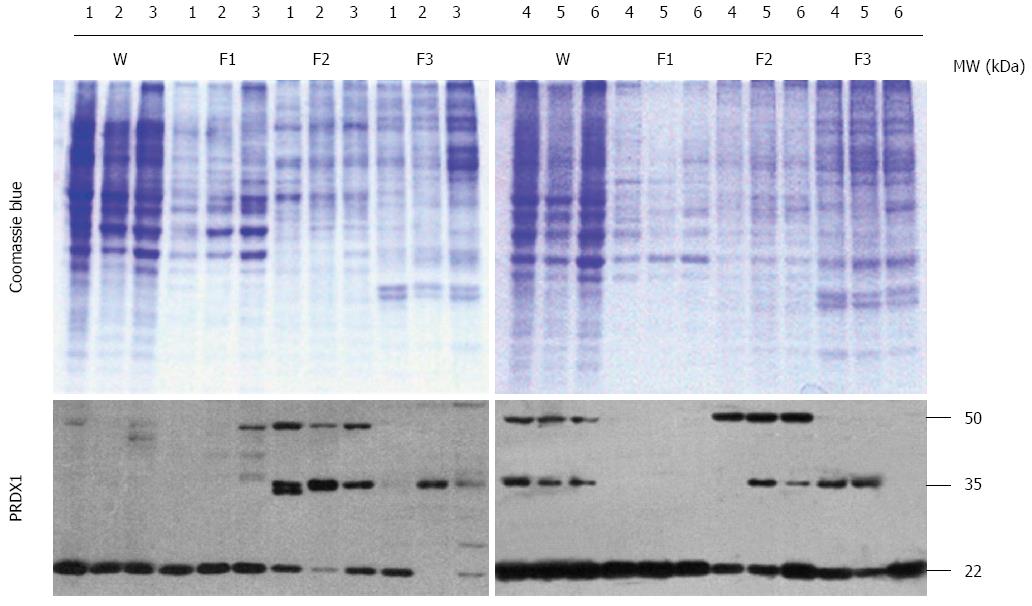
Figure 2 Western blotting analysis of peroxiredoxin 1 protein in liver cancer cells.
The codes of liver cancer cells: 1: HepG2; 2: Hep3B; 3: SK-HEP-1; 4: Bel-7404; 5: SMMC-7721; 6: HLE. W: Whole lysates; F1: Cytosol fraction; F2: Membrane/organelle fraction; F3: Nucleus fraction. The upper panel is the Coomassie Blue stained SDS-PAGE gel, and the lower panel is the Western blotting of PRDX1. PRDX1: Peroxiredoxin 1.
- Citation: Sun YL, Cai JQ, Liu F, Bi XY, Zhou LP, Zhao XH. Aberrant expression of peroxiredoxin 1 and its clinical implications in liver cancer. World J Gastroenterol 2015; 21(38): 10840-10852
- URL: https://www.wjgnet.com/1007-9327/full/v21/i38/10840.htm
- DOI: https://dx.doi.org/10.3748/wjg.v21.i38.10840


