Copyright
©The Author(s) 2015.
World J Gastroenterol. Sep 28, 2015; 21(36): 10427-10434
Published online Sep 28, 2015. doi: 10.3748/wjg.v21.i36.10427
Published online Sep 28, 2015. doi: 10.3748/wjg.v21.i36.10427
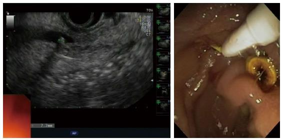
Figure 2 Endoscopic ultrasonography findings of common bile duct stone of 2.
2 mm in a nondilated common bile duct (left); common bile duct stone extraction during endoscopic retrograde cholangiopancreatography (right).
- Citation: Anderloni A, Galeazzi M, Ballarè M, Pagliarulo M, Orsello M, Piano MD, Repici A. Early endoscopic ultrasonography in acute biliary pancreatitis: A prospective pilot study. World J Gastroenterol 2015; 21(36): 10427-10434
- URL: https://www.wjgnet.com/1007-9327/full/v21/i36/10427.htm
- DOI: https://dx.doi.org/10.3748/wjg.v21.i36.10427


