Copyright
©The Author(s) 2015.
World J Gastroenterol. Sep 28, 2015; 21(36): 10367-10374
Published online Sep 28, 2015. doi: 10.3748/wjg.v21.i36.10367
Published online Sep 28, 2015. doi: 10.3748/wjg.v21.i36.10367
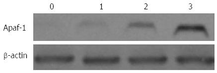
Figure 5 Ursodeoxycholic acid induces expression of APAF1.
Western blot analysis for the expression of APAF1 was performed with protein lysates prepared from control and treated xenografts. 0: control; 1-3 lanes: ursodeoxycholic acid (UDCA) 30 mg/kg per day, 50 mg/kg per day, and UDCA 70 mg/kg per day. β-actin was used as an internal control.
- Citation: Liu H, Xu HW, Zhang YZ, Huang Y, Han GQ, Liang TJ, Wei LL, Qin CY, Qin CK. Ursodeoxycholic acid induces apoptosis in hepatocellular carcinoma xenografts in mice. World J Gastroenterol 2015; 21(36): 10367-10374
- URL: https://www.wjgnet.com/1007-9327/full/v21/i36/10367.htm
- DOI: https://dx.doi.org/10.3748/wjg.v21.i36.10367


