Copyright
©The Author(s) 2015.
World J Gastroenterol. Sep 14, 2015; 21(34): 9936-9944
Published online Sep 14, 2015. doi: 10.3748/wjg.v21.i34.9936
Published online Sep 14, 2015. doi: 10.3748/wjg.v21.i34.9936
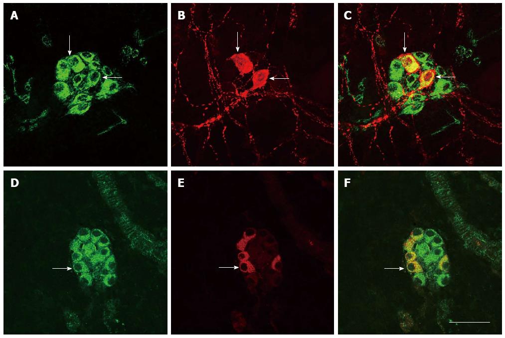
Figure 3 Immunofluorescence histochemical double-staining of the whole-mount sections of the rat mid-colon.
A and D: The endomorphin-2 (EM-2)-immunoreactive (IR) neurons; B and E: Vasoactive intestinal peptide (VIP)-IR neurons (B) and nitric oxide synthetase (NOS)-IR neurons (E) in the submucosal layer; C and F: Merged images of (A, B) and (D, E). Arrows point to neurons containing both EM-2 and VIP or NOS. Scale bar = 45 μm.
- Citation: Li JP, Zhang T, Gao CJ, Kou ZZ, Jiao XW, Zhang LX, Wu ZY, He ZY, Li YQ. Neurochemical features of endomorphin-2-containing neurons in the submucosal plexus of the rat colon. World J Gastroenterol 2015; 21(34): 9936-9944
- URL: https://www.wjgnet.com/1007-9327/full/v21/i34/9936.htm
- DOI: https://dx.doi.org/10.3748/wjg.v21.i34.9936


