Copyright
©The Author(s) 2015.
World J Gastroenterol. Sep 14, 2015; 21(34): 9887-9899
Published online Sep 14, 2015. doi: 10.3748/wjg.v21.i34.9887
Published online Sep 14, 2015. doi: 10.3748/wjg.v21.i34.9887
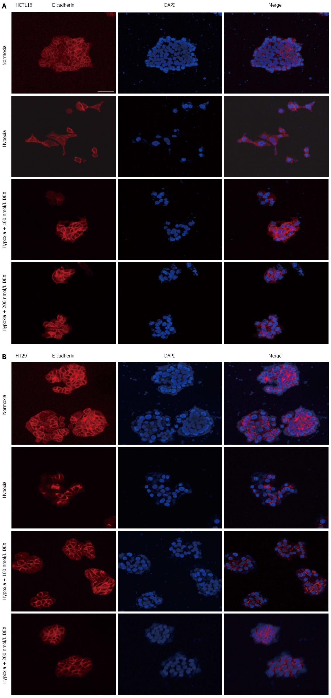
Figure 3 Effects of dexamethasone on cell morphology in hypoxia.
Immunocytochemical staining for E-cadherin and DAPI in HCT116 (A) and HT29 (B) cells. Cells were treated with DEX (100, 200 nmol/L) and/or hypoxia for 5 (A) or 7 d (B). E-cadherin (red) and DAPI (blue) were observed by immunocytochemistry. DEX rescued E-cadherin expression and morphological changes of HCT116 and HT29 cells under hypoxia. Scale bar, 50 μm; Magnification × 200. DEX: Dexamethasone; DAPI: 4',6-diamidino-2-phenylindole.
- Citation: Kim JH, Hwang YJ, Han SH, Lee YE, Kim S, Kim YJ, Cho JH, Kwon KA, Kim JH, Kim SH. Dexamethasone inhibits hypoxia-induced epithelial-mesenchymal transition in colon cancer. World J Gastroenterol 2015; 21(34): 9887-9899
- URL: https://www.wjgnet.com/1007-9327/full/v21/i34/9887.htm
- DOI: https://dx.doi.org/10.3748/wjg.v21.i34.9887


