Copyright
©The Author(s) 2015.
World J Gastroenterol. Sep 14, 2015; 21(34): 10045-10048
Published online Sep 14, 2015. doi: 10.3748/wjg.v21.i34.10045
Published online Sep 14, 2015. doi: 10.3748/wjg.v21.i34.10045
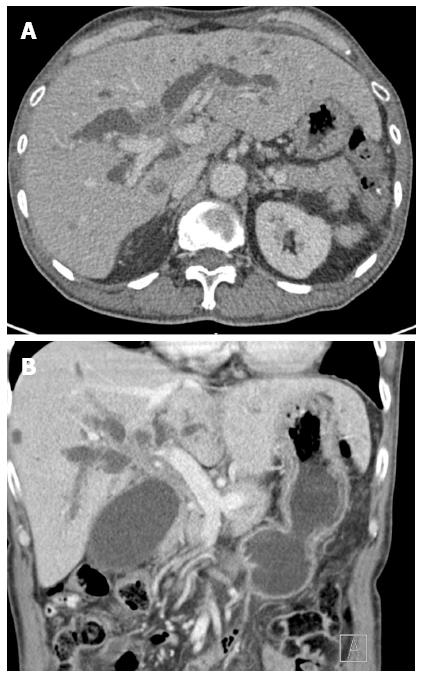
Figure 1 Computed tomography examination.
A: Axial; B: Coronal computed tomography images showing an ill-defined mass in the hilar region causing bilateral intrahepatic duct dilation.
-
Citation: Prachayakul V, Aswakul P. Endoscopic ultrasound-guided biliary drainage: Bilateral systems drainage
via left duct approach. World J Gastroenterol 2015; 21(34): 10045-10048 - URL: https://www.wjgnet.com/1007-9327/full/v21/i34/10045.htm
- DOI: https://dx.doi.org/10.3748/wjg.v21.i34.10045


