Copyright
©The Author(s) 2015.
World J Gastroenterol. Sep 7, 2015; 21(33): 9827-9832
Published online Sep 7, 2015. doi: 10.3748/wjg.v21.i33.9827
Published online Sep 7, 2015. doi: 10.3748/wjg.v21.i33.9827
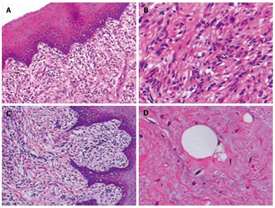
Figure 4 Histological analysis reveals a well-differentiated myxoid esophageal liposarcoma with a dedifferentiated component.
Hematoxylin and eosin stain of paraffin-embedded sections highlighting histological features of the case. A: Myxoid tumor cells located just under normal squamous cell epithelia (magnification × 100); B: Spindle tumor cells (magnification × 200); C: Dedifferentiated component of the liposarcoma (magnification × 100); D: Lipoblast cells (magnification × 200).
- Citation: Lin ZC, Chang XZ, Huang XF, Zhang CL, Yu GS, Wu SY, Ye M, He JX. Giant liposarcoma of the esophagus: A case report. World J Gastroenterol 2015; 21(33): 9827-9832
- URL: https://www.wjgnet.com/1007-9327/full/v21/i33/9827.htm
- DOI: https://dx.doi.org/10.3748/wjg.v21.i33.9827


