Copyright
©The Author(s) 2015.
World J Gastroenterol. Sep 7, 2015; 21(33): 9827-9832
Published online Sep 7, 2015. doi: 10.3748/wjg.v21.i33.9827
Published online Sep 7, 2015. doi: 10.3748/wjg.v21.i33.9827
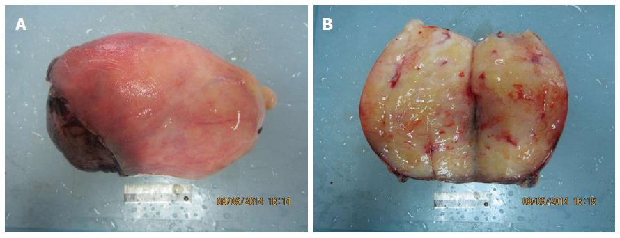
Figure 3 Surgical specimen of the esophageal liposarcoma.
Normal esophageal mucosa is shown covering the surface of the tumor. A yellow lobulated transmural mass invading the esophageal wall is visible at one surgical margin of the specimen.
- Citation: Lin ZC, Chang XZ, Huang XF, Zhang CL, Yu GS, Wu SY, Ye M, He JX. Giant liposarcoma of the esophagus: A case report. World J Gastroenterol 2015; 21(33): 9827-9832
- URL: https://www.wjgnet.com/1007-9327/full/v21/i33/9827.htm
- DOI: https://dx.doi.org/10.3748/wjg.v21.i33.9827


