Copyright
©The Author(s) 2015.
World J Gastroenterol. Sep 7, 2015; 21(33): 9808-9816
Published online Sep 7, 2015. doi: 10.3748/wjg.v21.i33.9808
Published online Sep 7, 2015. doi: 10.3748/wjg.v21.i33.9808
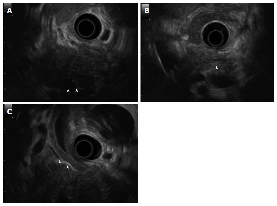
Figure 3 Findings of endoscopic ultrasonography.
Endoscopic ultrasonography revealed hypoechoic swelling of the pancreatic head (A: arrowheads), mild dilation of the upstream main pancreatic duct (B: arrowheads), and diffuse thickness of the common bile duct wall (C: arrowheads).
- Citation: Nakano E, Kanno A, Masamune A, Yoshida N, Hongo S, Miura S, Takikawa T, Hamada S, Kume K, Kikuta K, Hirota M, Nakayama K, Fujishima F, Shimosegawa T. IgG4-unrelated type 1 autoimmune pancreatitis. World J Gastroenterol 2015; 21(33): 9808-9816
- URL: https://www.wjgnet.com/1007-9327/full/v21/i33/9808.htm
- DOI: https://dx.doi.org/10.3748/wjg.v21.i33.9808


