Copyright
©The Author(s) 2015.
World J Gastroenterol. Sep 7, 2015; 21(33): 9793-9802
Published online Sep 7, 2015. doi: 10.3748/wjg.v21.i33.9793
Published online Sep 7, 2015. doi: 10.3748/wjg.v21.i33.9793
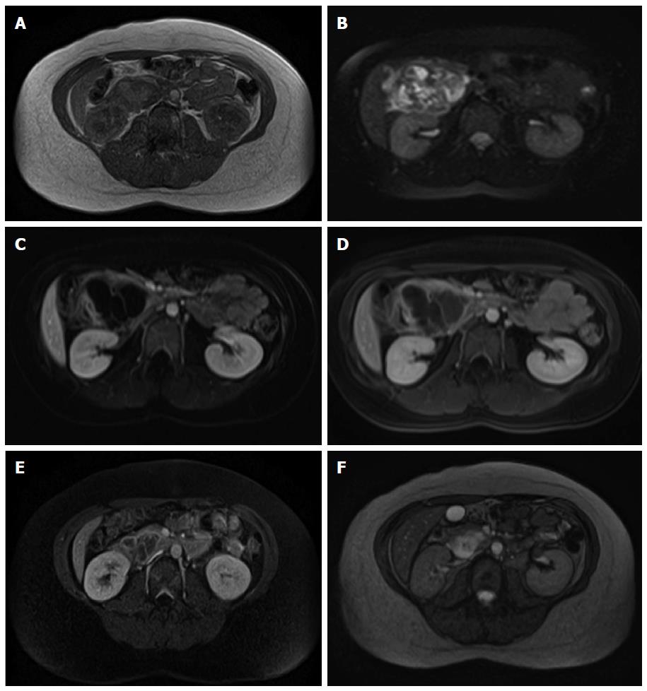
Figure 2 Magnetic resonance imaging.
Variable intensity in T1 weighted (A) and T2 weighted (B) imaging. Overall, the hemangioma had hypo-intensity in T1W and hyper-intensity in T2W imaging. Peripheral enhancement after gadolinium injection with gradual centripetal filling-in (C, D: Arterial phase; E: Venous; and F: Delayed venous phase) was noticed. Intensity was parallel to that of aorta. A central cystic area remained hypo-intense.
- Citation: Mondal U, Henkes N, Henkes D, Rosenkranz L. Cavernous hemangioma of adult pancreas: A case report and literature review. World J Gastroenterol 2015; 21(33): 9793-9802
- URL: https://www.wjgnet.com/1007-9327/full/v21/i33/9793.htm
- DOI: https://dx.doi.org/10.3748/wjg.v21.i33.9793


